Cranial & Brain Anatomy
The Skull
The skull is made up of 22 bones: the cranium includes eight bones that surround and protect the brain and 14 bones that form the face. In adults, all but one of the skull bones are fused together by immovable joints called sutures. The sutures lock the edges of the skull bones together, like pieces in a puzzle, to form a structure that is both rigid and strong. The mandible, or lower jaw, the only moveable skull bone, allows the mouth to open and close.


In newborns, the skull bones are not completely fused; they are linked by fibrous membrane called fontanelles. Fontanelles allow the skull to be compressed slightly during birth and accommodate growth of the brain during early infancy. By one-and-a-half years of age, the skull sutures have formed and the fontanels have disappeared.


Cranial Bones
The frontal bone forms the forehead. Two parietal bones form the sides of the cranial roof. Two temporal bones form the lower cranial sides. The occipital bone forms the cranial rear and floor. The ethmoid bone forms part of the nasal cavity. Shaped like a butterfly, the sphenoid bone forms the middle part of the cranial floor.
Facial Bones
The 14 facial bones provide the structure of the face and form the openings through which food, water, and air enter the body. Each of the following facial bones are paired: the maxillae form the upper jaw and front of the hard palate; the zygomatic bones form the cheeks; the nasal bones form the bridge of the nose; the lacrimal bones form part of the orbit, or eye socket; the palatine bones form the rear of the hard palate; and the inferior nasal conchae divide the nasal cavity. The vomer is a single bone that makes up part of the nasal septum, which divides the nostrils, and the mandible forms the lower jaw. The maxillae and mandible secure the teeth.


Small holes in the skull bones, called foraminae, and canals enable blood vessels, such as the carotid arteries and nerves, to enter and leave the skull. The spinal cord passes through a largest hole, called the foramen magnum, in the base of the cranium to join the brain. The occipital condyles on either side of the foramen magnum articulate with the first vertebra
(C1) of the spine to permit up-and-down movement of the head.
The Nervous System
The nervous system is a communication network that controls and coordinates most body actions. People are not generally conscious of some of these activities as they occur, such as regulating body temperature, breathing, or heart rate. Others, we consciously control, such as movement, talking, eating, and thinking.
The nervous system has two main parts: the central and the peripheral nervous system. The brain and spinal cord make up the central nervous system, or CNS. The nerves branching off the central nervous system make up the peripheral nervous system, or PNS. The PNS consists of nerves bundles made up of sensory and motor neurons.


Neurons
The nervous system is made up of cells called neurons. Neurons are long, thin cells that transmit electrical impulses. Neurons have many branched endings, called dendrites, which receive impulses from other neurons. An axon, or nerve fiber, carries nerve impulses to other neurons or to muscle. Neurons do not touch, but are separated by a tiny gap called a synapse. When an impulse arrives at the end of an axon, it releases chemicals that generate an impulse in the dendrites of the neighboring neuron.

There are three types of neurons: sensory, motor, and association. Sensory neurons transmit nerve impulses from sense organs (eyes, ears, nose, tongue, and touch) to the brain. They also carry nerve impulses to the brain and spinal cord. Motor neurons transmit nerve impulses from the brain and spinal cord to a specific area of the body. A nerve impulse to a muscle, for example, may cause it to contract. Association neurons make up 90 percent of all neurons and are found only in the brain and spinal cord.


Nerves
Nerves consist of bundles of both sensory and motor neurons.
Brain
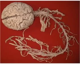
The brain is the control center of the nervous system. It enables us to think, feel, and move. The brain constantly receives information and sends out instructions to the body through the spinal cord and the body's vast network of nerves.


There are 12 pairs of cranial nerves branching off the brain. These nerves relay impulses from the sensory organs, such as the eyes or ears. Thirty-one pairs of spinal nerves branch off the spinal cord, exiting between each level of vertebrae. These nerves relay impulses to and from the rest of the body.


The largest part of the brain is the cerebrum, which controls the most sophisticated functions, such as thought, imagination, memory, emotion, speech, and sensory perception. The human cerebrum is quite large. It has two halves, or hemispheres. A band of nerve tissue, called the corpus callosum, links two halves to allow them to exchange information. Each hemisphere is covered by a layer of gray tissue, called the cerebral cortex, which is responsible for the higher functions of the brain, including conscious thought. The cortex is composed of sulci (folds) and gyri (bulges), which together provide a large surface area in the limited space inside the skull.

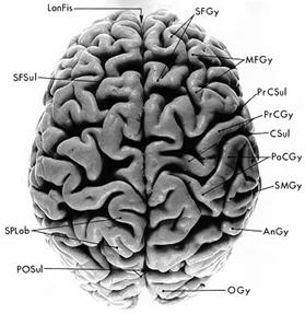
The cortex of each hemisphere has four lobes: the occipital, temporal, parietal, and frontal lobes. The occipital lobe controls vision. The temporal lobe controls sound and speech. The parietal lobe controls movement, touch, and recognition. And the frontal lobe controls thinking and planning.


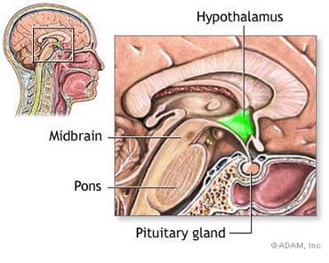
The brain stem and hypothalamus control automatic processes, such as breathing and heartbeat.


The cerebellum acts as a "mini brain" that coordinates body balance, posture, and movement.


Brain tissue is soft and delicate, and requires protection. In addition to the protection provided by the skull, the brain is surrounded by: the connective tissue membranes, or meninges; the dura mater; the arachnoid layer; and the pia mater. Beneath the arachnoid matter is a wide space filled with cerebrospinal fluid (CSF).
This fluid forms a liquid cushion that reduces the weight of the brain and protects it from knocks and jolts. CSF also helps the brain to receive nourishment.
Choose from the following categories to learn more about them: